Microsporomyces pseudomagnisporus Q.M. Wang, F.Y. Bai & A.H. Li 2020
MycoBank MB828820.
Holotype: China, Fanjingshan Mountain, Guizhou province, obtained from a leaf of an unidentified plant, Oct. 2011, Q.-M. Wang (holotype CGMCC 2.4538T preserved in a metabolically inactive state, ex-type CBS 15746 = FJS25C3).
Morphological description
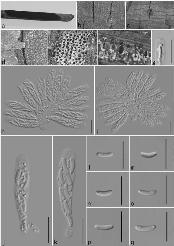
Culture characteristics: In YM broth, after 7 d at 17 °C, cells are cylindrical, 2.0–3.0 × 4.0–8.0 μm and single, budding is polar (Fig. 15I), a sediment is formed. After 1 mo at 17 °C, a part ring and sediment are present. On YM agar, after 1 mo at 17 °C, the streak culture is orange, butyrous, wrinkled and semi-glossy. The margin is entire. In Dalmau plate culture on corn meal agar, pseudohyphae are not formed. Sexual structures are not observed on YM, PDA, V8 and CM agar. Ballistoconidia are allantoid or reniform, 2.5–3.3 × 5.8–8.3 μm (Fig. 15J).
Habitat: A leaf of an unidentified plant.
Distribution: In China.
GenBank Accession: 18S+ITS+D1/D2: MK050384; RPB1: MK849125; RPB2: MK849351; EF1: MK849077
Notes:
Reference: A.-H. Li1,2, F.-X. Yuan1,3, M. Groenewald4 et al.
Vegetative cells grown in YM broth for 5 d at 17 °C and ballistoconidia produced on corn meal agar after 7 d at 17 °C. (A, B) Phy. aceris CGMCC 2.2662T; (C) Phy. jiayinensis CGMCC 2.5669T; (D) Me. layueensis CGMCC 2.5818T; (E) Sa. melibiophila CBS 5143T; (F) Mi. ellipsoideus CGMCC 2.5664T; (G, H) Mi. rubellus CGMCC 2.4444T; (I, J) Mi. pseudomagnisporus CGMCC 2.4538T; (K, L) Sy. rhododendri CGMCC 2.2613T; (M) Cy. raffinophilum CGMCC 2.3822T; (N) Cy. terricola CGMCC 2.3823T, (O) Do.ningxiaensis CGMCC 2.4451T; (P) Beg. foliicola CGMCC 2.3164T. Bars = 10 μm.