Serusiauxiella sinensis S.H. Jiang, Lücking & J.C. Wei, sp. nov. 2020
MycoBank MB 833570
Holotype: CHINA. Hainan: Ledong County, Jianfengling, Mingfenggu; 18° 44′ 32″ N, 108° 50′ 32″ E, 985 m; on living leaves; 12 December 2014, J.H. Wang & R.D. Liu HN2014281_2 (HMAS-L0141614).
Morphological description
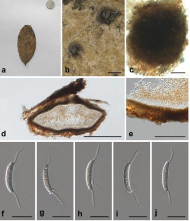
Thallus epiphyllous, subcuticular, 3–10 mm across and 10–20 μm thick, typically growing along the nerves, margins, and scars of leaves, circular, with entire to crenulate or lobulate margins, bright green but sometimes becoming whitish in the center. Photobiont a species of Trentepohlia, cells angular-rounded, 5–10 × 3–6 μm. Perithecia mostly erumpent, hemispherical to wart-shaped, 0.2–0.3 mm diam. and 100–180 μm high, exposed part black; excipulum prosoplectenchymatous, 5–10 μm thick, colorless to blackish brown; involucrellum carbonized, 10–30 μm thick, black; paraphyses unbranched or sparingly branched. Asci oblong, 40–65 × 5–10 μm. Ascospores biseriate or irregularly arranged, 8 per ascus, fusiform, with tapering, rounded to subacute ends, 1-septate, slightly constricted at the septum, not breaking into pieces, surrounded by a thin mucilaginous sheath, 10–18 × 3.5–5 μm. Pycnidia immersed-erumpent, wartshaped, those producing macroconidia 0.1–0.15 mm, those producing microconidia 0.05–0.1 mm diam., black. Macroconidia bacillar, 1-septate, 12–15 × 2–3.5 μm, with gelatinous appendages at both ends, 13–25 μm long but in squash mounts quickly growing to 70 μm or more. Microconidia fusiform, non-septate, 3–4 × 1–1.5 μm.
Habitat: on living leaves
Distribution: South China.
GenBank Accession: ITS MN720033
Notes: This new species is very similar to Serusiauxiella filifera and the only tangible difference appears to be the shorter asci. The differences in the ITS are, however, substantial, with an overall similarity of only 95.6% (see above).
Reference: Shu‑Hua Jiang , Robert Lücking , Amanda Barreto Xavier‑Leite3 et al.
Serusiauxiella sinensis sp. nov. (holotype, HMAS-L0141614) a Thallus, b Asci, c Ascospores, d Macroconidia showing the long-grown appendages. Scale bars: a = 600 μm, b–d = 10 μm