Dictyosporella guizhouensis J. Yang & K.D. Hyde 2020
Index Fungorum number: IF556314; Facesoffungi number: FoF 05999
Holotype: CHINA, Guizhou Province, Anshun City, Gaodang village, 26°4.267′ N, 105°41.883′ E, on decaying wood submerged in the Suoluo river, 19 October 2016, J. Yang, GD 34–2 (MFLU 18-1505, holotype; HKAS 102160, isotype).
Morphological description
Asexual morph: Colonies on natural substrate sporodochial, scattered, dark brown or black. Mycelium partly immersed, partly superficial, composed of septate, hyaline hyphae. Conidiophores macronematous, mononematous, compact, flexuous, simple or branched, mostly moniliform, with globose to subglobose, ellipsoid or clavate cells, hyaline, smooth-walled, up to 52 μm long. Conidiogenous cells monoblastic, integrated, terminal, globose, subglobose, ellipsoid or clavate, hyaline, smooth-walled, 9–17 × 6.5–12 μm ( x = 11.5 × 9.5 μm, n = 25). Conidia acrogenous, globose, subglobose, ellipsoid or irregular, muriform, with slightly irregular multi-euseptate, smooth, olivaceous to mid-brown, paler at the basal cell, 15–26 × 12.5–18.5 μm ( x = 19 × 16 μm, n = 50). Sexual morph: Undetermined.
Habitat: On decaying twigs from freshwater habitats.
Distribution: In China.
GenBank Accession: ITS: MK593606; LSU: MK593605; SSU: MK593611.
Notes: Dictyosporella, Junewangia and Sporidesmiella nested within Junewangiaceae in the phylogenetic tree based on a combined LSU and ITS sequence dataset (Fig. 64). However, Junewangia is shown to be polyphyletic which agrees with the result of Luo et al. (2019). D. guizhouensis clustered as a sister taxon to D. aquatica and D. thailandensis with full support (100% ML and 1.00 PP). D. hydei grouped with J. aquatica H.Y. Song & D.M. Hu, with full support (100% ML and 1.00 PP) and only one nucleotide difference, but only the LSU sequence data is available for D. hydei. Relationships between the genera and taxa in Junewangiaceae are awaiting to be resolved.
Reference: Hai‑Sheng Yuan1,2· Xu Lu1,2 · Yu‑Cheng Dai3 ·
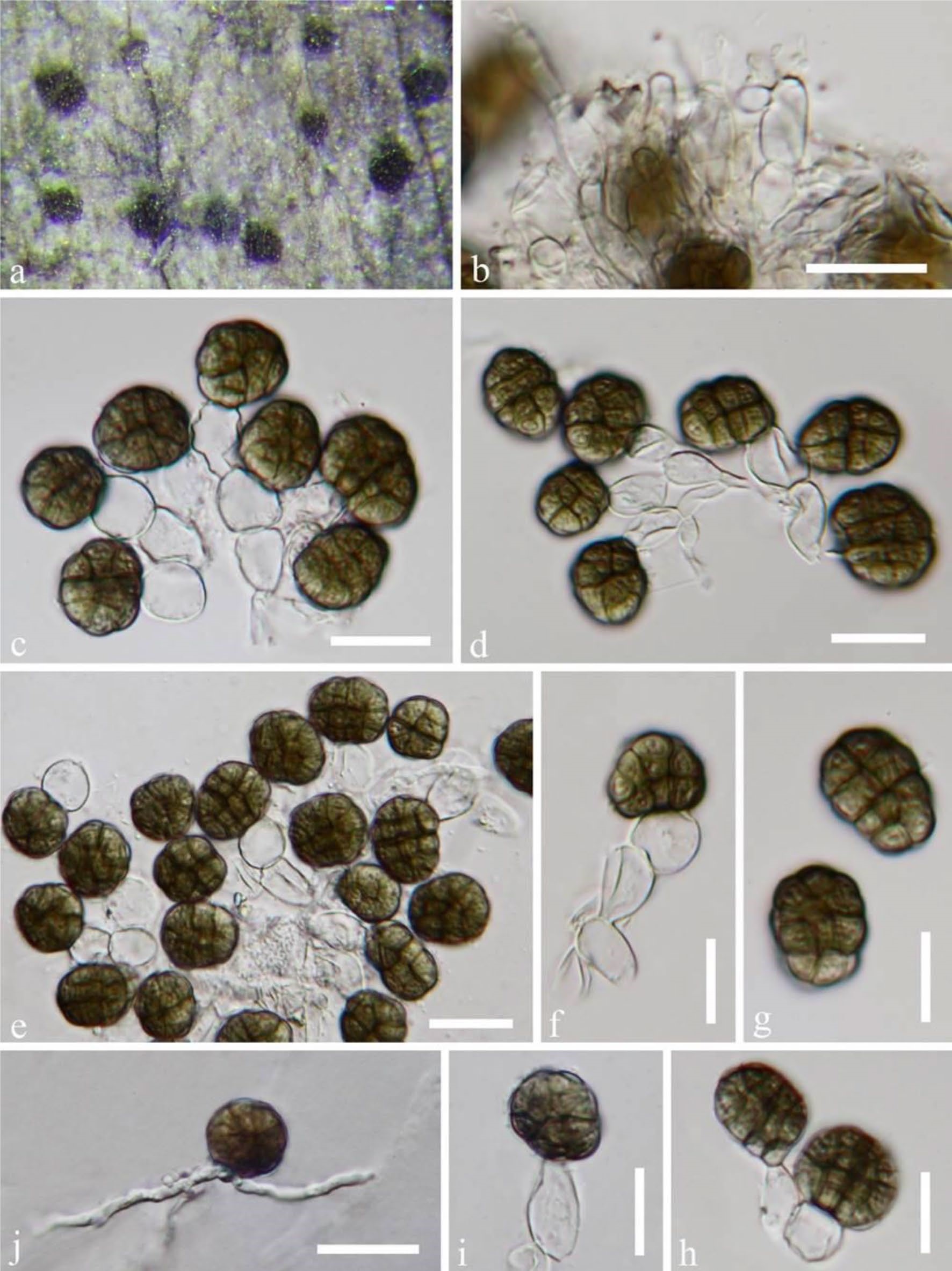
Dictyosporella guizhouensis (MFLU 18-1505, holotype). a Colony on substrate. b Conidiophores. c–f Conidiogenous cells with conidia. g–i Conidia. j Germinated conidium on PDA. Scale bars: b–e, j = 20 μm, f–i = 10 μm