Foraminispora austrosinensis (J.D. Zhao & L.W. Hsu) Y.F. Sun & B.K. Cui, comb. nov. 2020
MycoBank MB828440
Holotype: China, Hainan Province, Changjiang County, Yajia Forest Farm, on ground, 20 Apr. 1977, S.J. Han, HMAS 42695 (holotype, HMAS); Yunnan Province, Lincang County, 5 Aug. 2015, B.K. Cui, Cui 14318 (BJFC); ibid., Cui 14319 (BJFC). – Vietnam, Lam Dong Province, BidoupNuiba National Park, 15 Oct. 2017, B.K. Cui, Cui 16425 (BJFC).
Morphological description
Basidiomata annual, centrally to laterally stipitate, coriaceous to corky. Pileus single, suborbicular to umbelliform, up to 8.5 cm diam and 5 mm thick. Pileal surface yellow to yellowish brown when dry, dull, tomentose, with concentric furrows and radial wrinkles, sagging at the centre; margin acute to obtuse, entire, wavy and incurved when dry. Pore surface white to cream buff when fresh, colour unchanging when bruised, pale yellow when dry, even ferruginous in old specimens; pores circular to angular, 6–7 per mm; dissepiments slightly thick, entire. Context white to cream, without dark resinous lines, corky, up to 2 mm thick. Tubes concolorous with pore surface, hard corky, up to 3 mm long. Stipe concolorous with pileal surface, cylindrical and hollow, swollen at base, up to 7.5 cm long and 1 cm diam. Hyphal system trimitic; generative hyphae with clamp connections, all hyphae IKI–, CB+; tissues darkening in KOH.
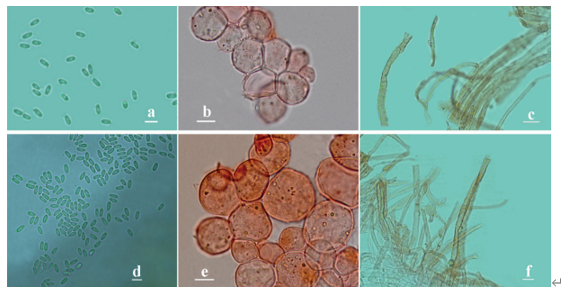
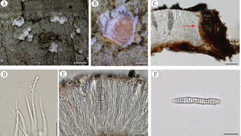
Generative hyphae in context colourless, thin-walled, 2–4 μm diam; skeletal hyphae in context colourless to pale yellow, thick-walled with a wide to narrow lumen or subsolid, arboriform branched and flexuous, 3–6 μm diam; binding hyphae in context colourless, subsolid, branched and flexuous, 1–2 μm diam. Generative hyphae in tubes colourless, thin-walled, 2–3 μm diam; skeletal hyphae in tubes colourless to pale yellow, thick-walled with a wide to narrow lumen or subsolid, arboriform branched and flexuous, 2–5 μm diam; binding hyphae in tubes colourless, subsolid, branched and flexuous, 1–2 μm diam. Pileal cover composed of clamped generative hyphae, thin- to thick-walled, apical cells clavate and inflated, often slanting to one side, yellowish brown, about 60–80 × 5–10 μm, forming a regular palisade. Cystidia absent; cystidioles clavate, colourless, thin-walled, 18–20 × 4–5 μm. Basidia barrel-shaped to clavate, colourless, thin-walled, 13–23 × 11–14 μm; basidioles in shape similar to basidia, colourless, thin-walled, 13–17 × 6–10 μm. Basidiospores globose to subglobose, pale yellow, IKI–, CB+, with double and distinctly thick walls, exospore wall smooth, endospore wall with conspicuous spinules, (7–)7.2– 8.5(–8.7) × (5.8–)6.7–8(–8.3) μm, L = 7.87 μm, W = 7.37 μm, Q = 1.06–1.07 (n = 60/2). Under SEM, exospore wall uneven or foveolate; endospore wall with some hollow and columnar spinules which persist to exospore wall forming holes.
Habitat: on ground
Distribution: Hainan Province, China.
GenBank Accession: ITS MK119809a; nLSU MK119888a; TEF MK121559a; TUB MK124987a
Notes: Amauroderma austrosinense was described from the tropical part of China (Zhao et al. 1984). It has the typical features such as white to straw colour pore surface when fresh and colour unchanging when bruised, cream context, hollow and columnar spinules on endospore wall which persist to the exospore wall forming the holes (Fig. 3a–b) that characterise Foraminispora (Costa-Rezende et al. 2017). In the phylogenetic analyses, A. austrosinense was shown as a distinct lineage in Foraminispora with high support (100 % ML, 1.00 BPP). Therefore, we transferred A. austrosinense to Foraminispora as a new combination.
Reference: Y.-F. Sun1,2, D.H. Costa-Rezende3, J.-H. Xing1 et al.
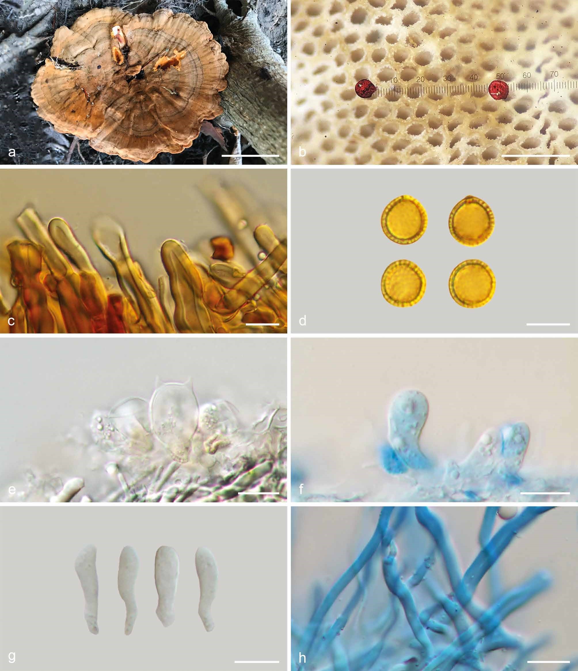
Basidiomata and microscopic structures of Foraminispora austrosinensis (Cui 16425). a. Basidiomata; b. pores; c. apical cells from pileal cover; d. basidiospores; e. basidia; f. basidioles; g. cystidioles; h. skeletal hyphae from context. — Scale bars: a = 2.5 cm; b = 0.5 mm; c, e–h = 10 µm; d = 7 µm.