Foraminispora concentrica (J. Song, Xiao L. He & B.K. Cui) Y.F. Sun & B.K. Cui, comb. nov. 2020
MycoBank MB828441
Holotype: China, Sichuan Province, Mianning County, Lingshansi Park, on ground of angiosperm forest, 13 Sept. 2015, B.K. Cui, Cui 12644 (holotype, BJFC); ibid., Cui 12646 (paratype, BJFC); ibid., Cui 12647 (paratype, BJFC); ibid., Cui 12648 (paratype, BJFC); Yunnan Province, Lanping County, Luoguqing, 18 Sept. 2017, B.K. Cui, Cui 16238 (BJFC); ibid., Cui 16239 (BJFC); ibid., Cui 16240 (BJFC); Guizhou Province, Liupanshui, Lingshansi Park, on stump of angiosperm tree, 15 Oct. 2018, H.J. Li, Cui 1510 (BJFC).
Morphological description
Basidiomata annual, centrally to laterally stipitate, coriaceous to corky. Pileus single, orbicular to suborbicular, up to 11 cm diam and 1 cm thick. Pileal surface yellowish brown to reddish brown, dull, tomentose, with obvious concentric zones and radial wrinkles, slightly sagging at the centre; margin acute, entire, faintly wavy and incurved when dry. Pore surface white. when fresh, colour unchanging when bruised, pale yellow to straw colour when dry; pores circular to angular, 3–5 per mm; dissepiments thin to slightly thick, entire. Context white to cream, without dark resinous lines, corky, up to 4 mm thick. Tubes concolorous with pore surface, hard corky, up to 6 mm long. Stipe concolorous with pileal surface, cylindrical and hollow, slightly swollen at base, up to 7.5 cm long and 6 mm diam. Hyphal system trimitic; generative hyphae with clamp connections, all hyphae IKI–, CB+; tissues darkening in KOH. Generative hyphae in context colourless, thin-walled, 3–4 μm diam, often collapsed; skeletal hyphae in context colourless to pale yellow, thick-walled with a wide to narrow lumen or subsolid, arboriform branched and flexuous, 2–6 μm diam; binding hyphae in context colourless, subsolid, branched and flexuous, 1–2 μm diam. Generative hyphae in tubes colourless, thin-walled, 2–3 μm diam; skeletal hyphae in tubes pale yellow, thick-walled with a wide to narrow lumen or subsolid, arboriform branched and flexuous, 2–5 μm diam; binding hyphae in tubes colourless, subsolid, branched and flexuous, 1–2 μm diam. Pileal cover composed of clamped generative hyphae, thin- to thick-walled, apical cells clavate, inflated or constricted, faintly slanting to one side, yellowish brown, about 30–60 × 6–11 μm, forming a regular palisade. Cystidia or cystidioles absent. Basidia barrel-shaped to clavate, colourless, thin-walled, 17–25 × 10–14 μm; basidioles in shape similar to basidia, colourless, thin-walled, 15–22 × 7–12 μm. Basidiospores subglobose to broadly ellipsoid, pale yellow, slightly dextrinoid, CB+, with double and distinctly thick walls, exospore wall smooth, endospore wall with conspicuous echinules, (6.8–)7.7–9.5(–9.6) × (6–)6.8–8.3(–9) μm, L = 8.6 μm, W = 7.48 μm, Q = 1.13–1.17 (n = 60/2). Under SEM, exospore wall uneven or foveolate, endospore wall with some hollow and columnar spinules which persist to exospore wall forming holes.
Habitat: on ground of angiosperm forest
Distribution: Sichuan Province, China.
GenBank Accession: ITS MK121499a; nLSU MK121561a; RPB2 MK119813a; TEF MK119892a
Notes: Amauroderma concentricum is distributed in temperate and subtropical areas of China. Based on its typical features and phylogenetic support, we transfer A. concentricum to Foraminispora as a new combination. Foraminispora concentrica is similar to Fo. austrosinensis by the yellow brown and tomentose concentrically zonate pileal surface, white context and pore surface, but the pores (6–7 per mm) and basidiospores (7.2–8.5 × 6.7–8 μm) of Fo. austrosinensis are relatively smaller than Fo. concentrica; moreover, the basidiospores of Fo. concentrica are slightly dextrinoid, while Fo. austro sinensis has non-dextrinoid basidiospores.
Reference: Y.-F. Sun1,2, D.H. Costa-Rezende3, J.-H. Xing1 et al.
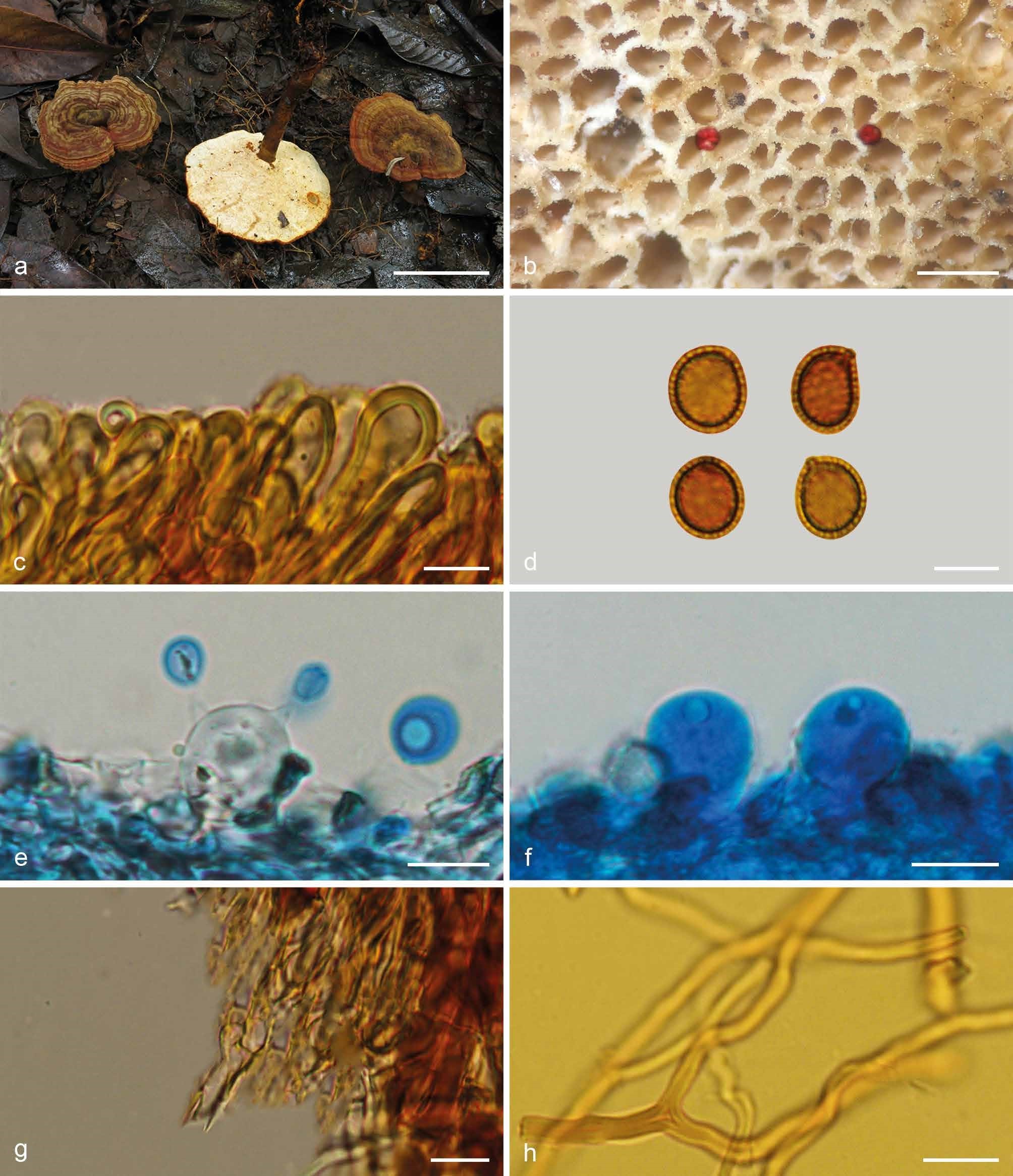
Basidiomata and microscopic structures of Foraminispora concentrica (Cui 12648). a. Basidiomata; b. pores; c. apical cells from pileal cover; d. basidiospores; e. basidia; f. basidioles; g. generative hyphae from context; h. skeletal hyphae from context. — Scale bars: a = 3 cm; b = 0.5 mm; c, e–h = 10 µm; d = 7 µm.