Tubakia dryinoides C. Nakash., Fungal Systematics and Evolution 1: 80 (2018)2021
MycoBank No:
Holotype:
Morphological description
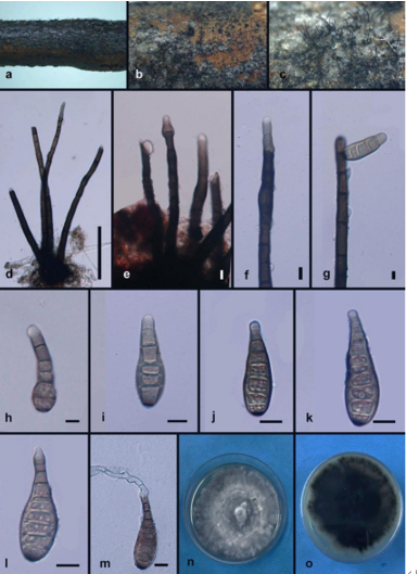
Asexual morph: Living as endophyte in leaves, forming distinct leaf lesions, shape and size variable, subcircular to angular-irregular, pale brown to brown. Colonies on PDA incubated at 25°C in the dark with an average radial growth rate of 5–7 mm/d and occupying an entire 90 mm Petri dish in 14 d, forming some conspicuous concentric circles, aerial mycelium cottony, white initially, then becoming greyish-sepia. Conidiomata sporodochial, appeared within 14 days or longer, formed on agar surface, slimy, black, semi-submerged. Sporodochial conidiophores densely and irregularly branched, 11.0–24.0 μm × 1.5–5.0 μm, bearing apical whorls of 2–3 phialides; sporodochial phialides monophialidic, subulate to subcylindrical, 9.0–16.0 μm × 1.5–5.0 μm, smooth, thin-walled, apex obtuse to truncate, sometimes forming indistinct periclinal thickenings. Conidia solitary, ellipsoid to obovoid, 6.5–14.0 μm × 4.0–6.0 μm, wall thin, up to 1.0 μm, hyaline to subhyaline, smooth, apex and base broadly rounded, with inconspicuous to conspicuous basal hilum (frill), occasionally somewhat peg-like and truncate when conspicuous. Microconidia not observed.
Sexual morph not observed
Culture characteristics. Cultures incubated on MEA at 25°C in darkness, attaining 38.0–42.0 mm diam. after 14 d (growth rate 2.7–3.0 mm diam./d), margin scal- loped, at first creamy white, grey near the centre, reverse light brown to dark, with olivaceous edge. Conidial formation not observed
Habitat: on diseased leaves of Quercus palustris (Fagaceae),
Distribution: China, Shandong Province: Zibo Lushan National Forest Park,
GenBank Accession: its MW784842; lsu MW784852; tef1 MW842260 ; tub2 MW842263 ; rpb2 MW842266.
Notes: Braun et al. (2018) described Tubakia dryinoides, based on morphological and molecular data. The holotype of T. dryinoides (NBRC H-11618) was collected from Quercus phillyraeoides A. Gray (Braun et al. 2018). In our current research, isolate (SAUCC 1924) collected from diseased leaves of Quercus palustris clustered in the Tubakia dryinoides clade by strong support (Figs. 1 and 2). We, therefore, consider the isolated strain (SAUCC 1924) as T. dryinoides. The conidiomata of T. dryinoides is only known from true pycnothyria and the sporodochial conidiomata of the isolated strain (SAUCC 1924) is new for T. dryinoides (Braun et al. 2018). Additionally, the conidia of our isolate (SAUCC 1924) is narrower than the original description of T. dryinoides (4.0–6.0 μm vs. 5.5–10.0 μm; Braun et al. 2018)
Reference: [1] Zhang, Z. , Mu, T. , Liu, S. , & Xia, J. W. . (2021). Morphological and phylogenetic analyses reveal a new genus and two new species of tubakiaceae from china. MycoKeys.
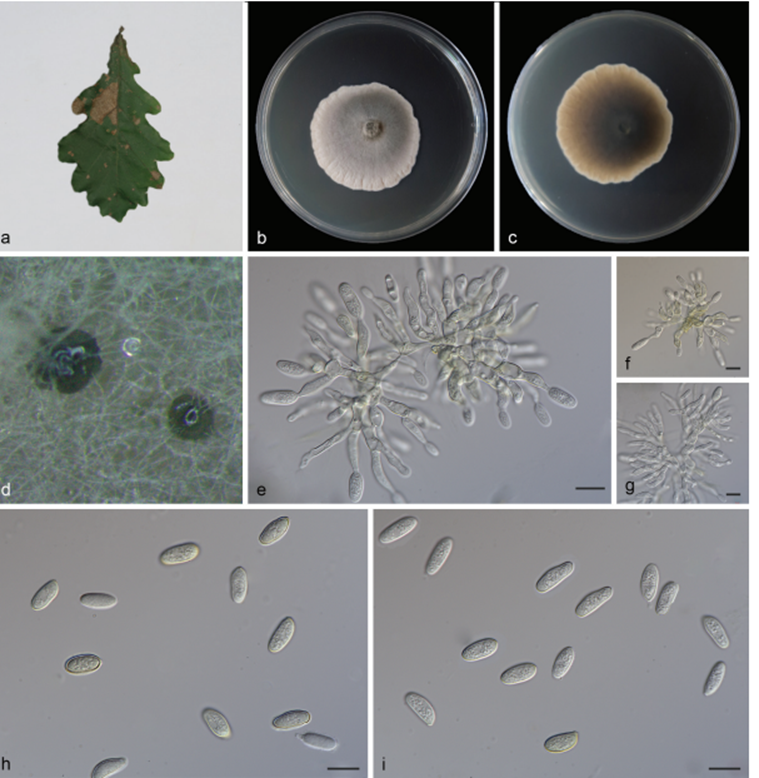 Tubakia dryinoides (SAUCC 1924). a diseased leaf of Quercus palustris; b surface of colony after 15 days on MEA; c reverse of colony after 15 days on MEA; d conidiomata; e–g conidiophores, conidiogenous cells with conidia; h–i conidia. Scale bars: 10 μm (e–i)