Tubakia lushanensis Z. X. Zhang, J. W. Xia & X. G. Zhang, sp. nov. 2021
MycoBank No: 841105
Holotype: HSAUP1923, ex-type living culture SAUCC 1923)
Morphological description
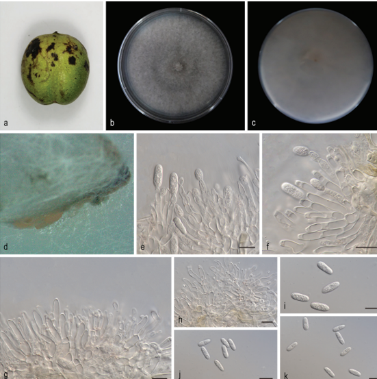
Asexual morph: : Leaf spots irregular, occurring on leaf veins and at leaf edges. Colonies on PDA incubated at 25°C in the dark with an average radial growth rate of 5–7 mm/d and occupying an entire 90 mm Petri dish in 14 d, forming some conspicuous concentric circles, aerial mycelium cottony, white initially, then becoming greyish-sepia. Conidiomata pycnidial, usually globose or subglobose when viewed from above, formed on agar surface, black, semi-submerged, up to 200 μm diam. Pycnidial wall composed of an outer layer of yellow-brown, thick-walled textura angularis and an inner layer with hyaline, thin-walled cells. Conidiophores reduced to conidiogenous cells lining the inner cavity, ampulliform or flask-shaped, smooth, hyaline, 9.0–15.0 μm × 2.0–4.0 μm. Conidia solitary, globose to irregular globose, ellipsoid to broad ellipsoid, 10.0–18.0 μm × 7.5–16.0 μm, length/width ratio 1.0–1.7, slightly lighter and wall thin when immature, slightly darker and wall thickened when ripening, smooth, apex rounded, base with peg-like hila, 1.3–2.3 μm diam. Microconidia not observed.
Sexual morph not observed.
Culture characteristics. Cultures incubated on MEA at 25°C in darkness, attaining 52.0–56.0 mm diam. after 14 d (growth rate 3.7–4.0 mm diam./d), creamy white to pale brown with regular margin, grey near the centre and hyphae clusters, reverse brown to dark brown rings, heterogeneous colour, with creamy-white edge. Conidial formation not observed.
Habitat: , on diseased leaves of Quercus palustris Münchh
Distribution: China, Shandong Province: Zibo Lushan National Forest Park
GenBank Accession: its MW784677; lsu MW784850 ; tef1 MW842262; tub2 MW842265;rpb2 MW84226.
Notes:
Reference: [1] Zhang, Z. , Mu, T. , Liu, S. , & Xia, J. W. . (2021). Morphological and phylogenetic analyses reveal a new genus and two new species of tubakiaceae from china. MycoKeys.
 Tubakia lushanensis (SAUCC 1923). a diseased leaf of Quercus palustris; b surface of colony after 15 days on MEA; c reverse of colony after 15 days on MEA; d conidiomata; e–i conidiogenous cells with conidia; j–k conidia. Scale bars: 10 μm (e–k).