Foraminispora yunnanensis (J.D. Zhao & X.Q. Zhang) Y.F. Sun & B.K. Cui, comb. nov. 2020
MycoBank MB MB828442
Holotype: China, Yunnan Province, Xichou County, Xiaoqiaogou, on ground, 14 May 1959, Q.Z. Wang, HMAS 48231 (holotype, HMAS); Kunming, Qiongzhusi Park, on ground of angiosperm forest, 21 Oct. 2009, B.K. Cui, Cui 7974 (BJFC).
Morphological description
Basidiomata annual, centrally to laterally stipitate, coriaceous to corky. Pileus single, suborbicular to flabelliform, up to 6.5 cm diam and 6 mm thick. Pileal surface cinnamon to reddish brown when dry, dull, tomentose, with faintly concentric furrows at the margin and irregular wrinkles; margin subacute to obtuse, entire and wavy when dry. Pore surface white to pale straw, colour unchanging when bruised; pores circular to angular, 2–3 per mm; dissepiments thin, fragile when dry. Context white to pale yellow, without dark resinous lines, corky, up to 2 mm thick. Tubes concolorous with pore surface, occasionally fascicular, woody hard, up to 4 mm long. Stipe concolorous with pileal surface, cylindrical and solid, slightly swollen at base, up to 8 cm long and 6 mm diam. Hyphal system trimitic; generative hyphae with clamp connections, all hyphae IKI–, CB+; tissues darkening in KOH. Generative hyphae in context colourless, thin-walled, 3–4 μm diam; skeletal hyphae in context pale yellow, thick-walled with a wide to narrow lumen or subsolid, arboriform branched and flexuous, 3–6 μm diam; binding hyphae in context colourless, subsolid, branched and flexuous, 1–3 μm diam. Generative hyphae in tubes colourless, thin-walled, 2–4 μm diam; skeletal hyphae in tubes colourless to pale yellow, thick-walled with a wide to narrow lumen or subsolid, arboriform branched and flexuous, 2–5 μm diam; binding hyphae in tubes colourless, subsolid, branched and flexuous, 1–2 μm diam. Pileal cover composed of clamped generative hyphae, thin- to thick-walled, apical cells clavate, inflated or constricted, arranged loosely, yellowish brown, about 40–60 × 6–9 μm, forming an irregular palisade. Cystidia or cystidioles absent. Basidia barrel-shaped to clavate, colourless, thin-walled, 20–35 × 11–18 μm; basidioles in shape similar to basidia, colourless, thin-walled, 13–23 × 6–10 μm. Basidiospores broadly ellipsoid to ellipsoid, pale yellow, IKI–, CB+, with double and distinctly thick walls, exospore wall smooth, endospore wall with conspicuous spinules, 8–10.7(–11.2) × (6.8–)7–8.3(–8.7) μm, L = 9.35 μm, W = 7.63 μm, Q = 1.18–1.27 (n = 60/2). Under SEM, exospore wall uneven or foveolate, endospore wall with some hollow and columnar spinules which persist to exospore wall forming holes.
Habitat: on ground
Distribution: Yunnan Province, China.
GenBank Accession: ITS KJ531653; nLSU KU220013
Notes: Foraminispora yunnanensis can be distinguished by large lacerate pores (2–3 per mm), occasionally fascicular tubes and broadly ellipsoid to ellipsoid basidiospores. It is also consistent with Foraminispora in the unique ultrastructural characteristics of the exospore wall with obvious holes caused by hollow and columnar spinules on the endospore wall which persist to the exospore wall (Fig. 3g–h). In the phylogenetic analyses, Fo. yunnanensis was shown to be a distinct lineage in Foraminispora with high support (100 % ML, 1.00 BPP).
Reference: Y.-F. Sun1,2, D.H. Costa-Rezende3, J.-H. Xing1 et al.
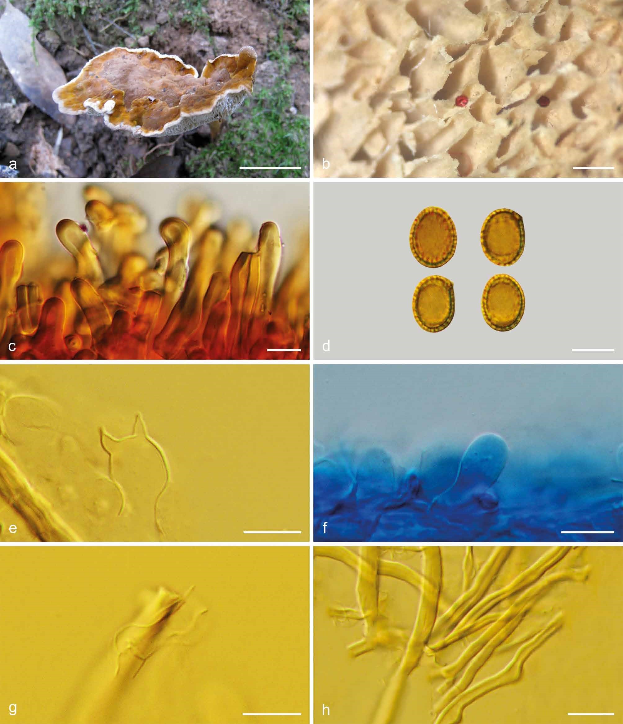
Basidiomata and microscopic structures of Foraminispora yunnanensis (Cui 7974). a. Basidiomata; b. pores; c. apical cells from pileal cover; d. basidiospores; e. basidia; f. basidioles; g. generative hyphae from context; h. skeletal hyphae from tubes. — Scale bars: a = 2 cm; b = 0.5 mm; c, e–h = 10 µm; d = 7 µm.