Sanguinoderma bataanense (Murrill) Y.F. Sun & B.K. Cui, comb. nov. 2020
MycoBank MB828443
Holotype: China, Hainan Province, Danzhou, on stump of Litchi chinensis, 7 May 2009, B.K. Cui, Cui 6285 (BJFC); ibid., Y.C. Dai, Dai 10746 (BJFC); Guangxi Autonomous Region, Baise, Jinzhongshan Nature Reserve, on rotten wood, 21 Oct. 1957, L.W. Xu, HMAS 23492 (HMAS).
Morphological description
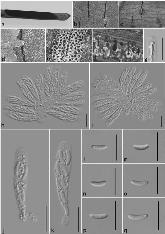
Basidiomata annual, laterally stipitate, soft to hard corky. Pileus single, suborbicular to flabelliform, up to 3.5 cm diam and 8 mm thick. Pileal surface dark yellowish brown when dry, dull, glabrous, with concentric furrows and radial wrinkles; margin obtuse, entire, wavy and incurved when dry. Pore surface grey- ish white when fresh, colour changing to blood red when bruised, then quickly darkening; pores subcircular to irregular, 1–4 per mm; dissepiments thin, entire. Context dark brown, with thick black stratum, corky, up to 2 mm thick. Tubes darker than context, woody hard, up to 6 mm long. Stipe concolorous with pileal surface, cylindrical and hollow, slightly swollen at the base, up to 4.5 cm long and 3 mm diam. Hyphal system trimi tic; generative hyphae with clamp connections, all hyphae IKI–, CB+; tissues darkening in KOH. Generative hyphae in context colourless, thin-walled, 2–4 μm diam; skeletal hyphae in context pale yellow, thick-walled with a wide to narrow lumen or subsolid, arboriform branched and flexuous, 2–6 μm diam; binding hyphae in context pale yellow, subsolid, branched and flexuous, 1–2 μm diam. Generative hyphae in tubes colourless, thin-walled, 2–3 μm diam; skeletal hyphae in tubes pale yellow, thick-walled with a wide or narrow lumen to subsolid, arboriform branched and flexuous, up to 7 μm diam; binding hyphae in tubes pale yellow, subsolid, branched and flexuous, 1–2 μm diam. Pileal cover composed of clamped generative hyphae, thin- to thick-walled, apical cells clavate with obvious septa, slightly inflated, reddish brown, about 30–70 × 5–9 μm, forming a regular palisade. Cystidia or cystidioles absent. Basidia barrel-shaped to clavate, colourless, thin-walled, 30–40 × 12–16 μm; basidioles in shape similar to basidia, colourless, thin-walled, 25–30 × 8–13 μm. Basidiospores globose to subglobose, pale yellow, IKI–, CB+, with double and distinctly thick walls, exospore wall smooth, endospore wall with conspicuous spinules, (9.6–)10–12.5(–13.1) × (8.5–)9–11(–11.3) μm, L = 11.13 μm, W = 9.84 μm, Q = 1.13 (n = 60/2). Under SEM, exospore wall thin and badly worn, endospore wall with long and thick columnar spinules which are loosely arranged and cause conspicuous warts on exospore wall (Fig. 8a–b).
Habitat: on stump of Litchi chinensis
Distribution: China. Philippines.
GenBank Accession: ITS MK119831a; nLSU MK119910a; RPB2 MK121537a; TEF MK121580a; TUB MK124997a
Notes: Amauroderma bataanense was described from the Philippines (Murrill 1908). We collected it from the tropical areas of China. It can be distinguished by its fresh pore surface colour that changes to blood red when bruised, subcircular to irregular pores (1–4 per mm) with thin and entire dissepiments, and distinctly thick-walled basidiospores. In the phylogenetic analyses, A. bataanense was shown to be a distinct well-supported lineage in Sanguinoderma with strong support (Fig. 1, 2). Therefore, we transferred A. bataanense to Sanguinoderma.
Reference: Y.-F. Sun1,2, D.H. Costa-Rezende3, J.-H. Xing1 et al.
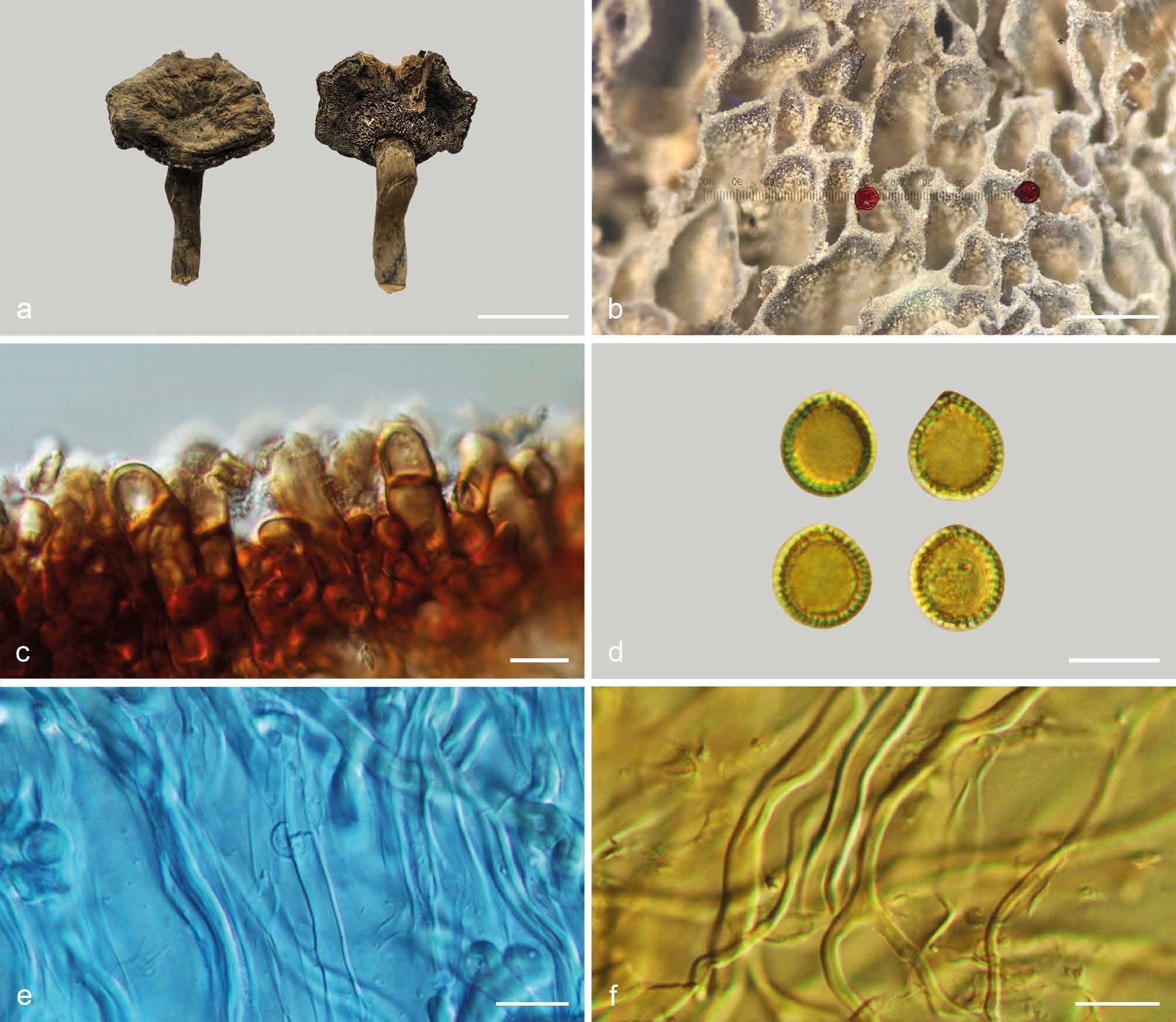
Basidiomata and microscopic structures of Sanguinoderma bataanense (Dai 10746). a. Basidiomata; b. pores; c. apical cells from pileal cover; d. basidiospores; e. generative hyphae from tubes; f. skeletal hyphae from context. — Scale