Sanguinoderma elmerianum (Murrill) Y.F. Sun & B.K. Cui, comb. nov. 2020
MycoBank MB828444
Holotype: China, Yunnan Province, Xishuangbanna, Baka Xiao Zhai Nature Reserve, 5 Aug. 2003, T.Z. Wei, HMAS 133187 (HMAS); Guangdong Province, Zhaoqing, Dinghushan Nature Reserve, dead angiosperm tree, 30 June 2010, B.K. Cui, Cui 8940 (BJFC).
Morphological description
Basidiomata annual, lateral stipitate to almost sessile, coriaceous to soft corky. Pileus single, flatly reniform, up to 14.5 cm diam and 1 cm thick. Pileal surface dark brown to nearly black, dull, glabrous, with concentric furrows and radial wrinkles; margin obtuse, entire, slightly wavy and flat when dry. Pore surface pale brown to pale grey when fresh, colour changing to blood red when bruised, then quickly darkening; pores cir- cular to angular, 5–7 per mm; dissepiments slightly thick, entire. Context pale brown to cinnamon, with two dark melanoid lines, soft corky, up to 4 mm thick. Tubes slightly darker than context, concolorous with pore surface, hard corky, up to 6 mm long. Stipe concolorous with pileal surface, cylindrical, swollen at base, about 9 cm long and 3 cm diam. Hyphal system trimitic; generative hyphae with clamp connections, binding hyphae usually serrated, all hyphae IKI–, CB+; tissues darkening in KOH. Generative hyphae in context colourless, thin-walled, 2–4 μm diam, usually collapsed; skeletal hyphae in context colourless to pale yellow, thick-walled with a wide or narrow lumen to subsolid, arboriform branched and flexuous, 3–6 μm diam; binding hyphae in context colourless, subsolid, branched and flexuous, 1–2 μm diam. Generative hyphae in tubes colourless, thin-walled, 2–4 μm diam; skeletal hyphae in tubes pale yellow, with a wide to narrow lumen or subsolid, arboriform branched and flexuous, 3–7 μm diam; binding hyphae in tubes colourless to pale yellow, subsolid, branched and flexuous, 1–2 μm diam. Pileal cover composed of clamped generative hyphae, thin- to thick-walled, apical cells clavate with obvious septa, inflated and slightly tortuous, pale brown to reddish brown, about 30–50 × 5–9 μm, forming a regular palisade. Cystidia or cystidioles absent. Basidia barrel-shaped to clavate, colourless, thin-walled, about 22–25 × 10–17 µm; basidioles in shape similar to basidia, colourless, thin-walled, 14–22 × 6–13 μm. Basidiospores globose to subglobose, pale yellow, IKI–, CB+, with double and medially thick walls, exospore wall smooth, endospore wall with conspicuous spinules, (8.4–)9.2–11.1(–11.2) × (7.8–)9–10.1(–10.5) μm, L = 10.26 μm, W = 9.5 μm, Q = 1.07–1.09 (n = 60/2). Under SEM, exospore wall alveolate to semi-reticulate, endospore wall with long and medially thick coniform spinules loosely arranged.
Habitat: unidentified
Distribution: China. Philippines.
GenBank Accession: ITS MK119833a; nLSU MK119912a
Notes: Amauroderma elmerianum was described from the Philippines (Murrill 1907). The specimens collected from China have soft basidiomata with a flat reniform pileus, and globose to subglobose basidiospores with an ornamented endospore wall (Fig. 8c–d); characters which are consistent with the previous descriptions (Murrill 1907, Furtado 1981). In the phylogenetic analyses, A. elmerianum was shown as a distinct well-supported lineage in Sanguinoderma with high support (100 % ML, 1.00 BPP). Therefore, we transferred A. elmer arium to Sanguinoderma.
Reference: Y.-F. Sun1,2, D.H. Costa-Rezende3, J.-H. Xing1 et al.
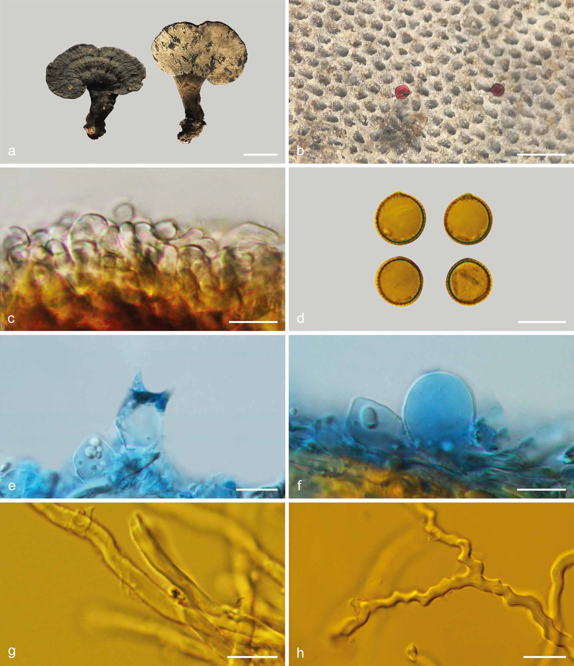
Basidiomata and microscopic structures of Sanguinoderma elmerianum (HMAS 133187). a. Basidiomata; b. pores; c. apical cells from pileal cover; d. basidiospores; e. basidia; f. basidioles; g. skeletal hyphae from tubes; h. binding hyphae from context. — Scale bars: a = 5 cm; b = 0.5 mm; c–h = 10 µm