Gymnosporangium lianhuaense Y.M. Liang, B. Cao & S.Q. Tao, sp. nov. 2020
MycoBank MB824626
Holotype: CHINA. SHAANXI: Baoji, Lianhua Mountain (34°12′04″N, 106°35′47″E; 2166 m asl), on Juniperus chinensis, 28 Apr 2017, S.Q. Tao BJFC- R02959 (holotype).
Morphological description
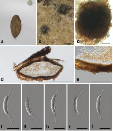
Spermogonia epiphyllous, aggregated to groups, pale yellow to black. Aecia hypophyllous, roestelioid, 1–6 mm long, peridium splitting at apex, retaining tubular form, peridial cells rhomboid, 48–105 × 14–32 μm, inner wall and side wall moderately rugose (type MR), outer wall smooth (type S). Aeciospores globoid, yellowish brown, 18–28 × 16–22 μm, wall 0.7–2 μm thick, surfy, more than 5 pores, scattered. Telia caulicolous, causing irregular and successive gall-like knots, up to 20 cm, sori breaking forth on galls, variable in shape. Teliospores 2-celled, ellipsoid, oblong or fusiform, 38–57 × 14–24 μm, wall 1–2.5 μm thick, slightly narrowed above and below, slightly constricted at septum, smooth, yellowish brown, 2 pores in each cell, most (70%) distal cells have 1 pore apical and 1 pore near the septum, the basal cell with 2 pores near the septum, pedicels colorless, very long.
Habitat: on Juniperus chinensis
Distribution: Shaanxi in China
GenBank Accession: ITS2 MH178642; 28S MH184490; tef1 MH202913.
Notes: The surface structure of aeciospores of G. lianhuaense differs from other reported ones, here defined as a new surface structure (type “surfy”) for the resemblance to ocean waves. Processes of this structure are spindrift-shaped and irregular-shaped, 0.4–0.8 μm in height and 0.6–2 μm in width. Heads of processes granulate with small papillae and basal parts of processes are jointed by narrow reticulate ridges. This type is similar to the type M, but smaller in the size of processes and different in the shape of their heads (FIG. 15B, C). The life cycle of this species has been identified based on homology of sequences obtained from aecial and telial stages.
Reference: Si-Qi Tao, Bin Cao, Makoto Kakishima et al. (2020) Species diversity, taxonomy, and phylogeny of Gymnosporangium in China.
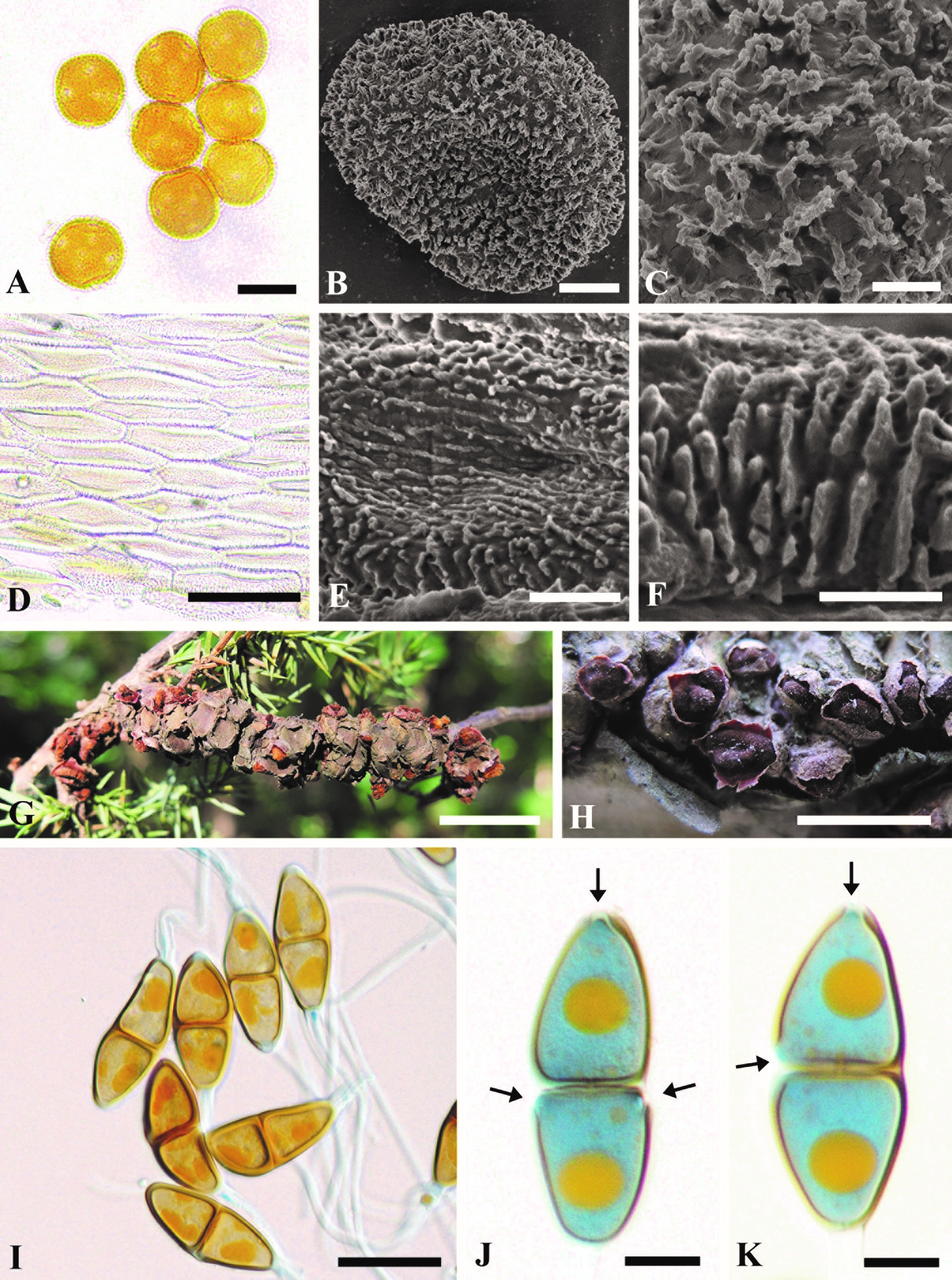
Morphology of Gymnosporangium lianhuaense. A. Aeciospores. B, C. Surface structure of aeciospores. D. Peridial cells. E. Inner wall of the peridial cells. F. Side wall of the peridial cells. G, H. Telia. I–K. Teliospores. Bars: A = 20 μm; B = 5 μm; C = 2 μm; D = 80 μm; E = 10 μm; F = 5 μm; G = 2 mm; H = 1 mm; I = 40 μm; J, K = 10 μm.